Female reproductive system - An easy understanding.

The female reproductive system is far more complex than the male because in addition to gamete formation, it has to nurture the developing foetus.
These parts
along with the mammary glands are integrated structurally and
functionally to support the,
1) Pair of Ovaries:
Ovarian stroma:
Outer cortex:
2) Accessory organs:
consists of:
1.
Fallopian tubes (Salpinges):
Location: In the female abdominal cavity. It extends from periphery of each ovary to uterus.
Size:
The edges of infundibulum have finger like projections called fimbriae (helps in collection of ova after ovulation).
Location: Proximal part of the fallopian tube.
Function: connects ampulla and infundibulum to uterus.
Size: 2 cm (0.8 inch) long.
2. Uterus (womb):
Location: Between urinary bladder and rectum, lying in pelvic cavity.
Perimetrium:
Endometrium:

The female reproductive system is far more complex than the male because in addition to gamete formation, it has to nurture the developing foetus.
It
consists of:
1) Pair of ovaries,
2) Accessory organs,
1. The fallopian tubes,
2. Uterus,
a) Fundus,
b) Body,
c) Cervix.
a) Fundus,
b) Body,
c) Cervix.
3. Vagina,
3) The external genitalia.
1. Mons Pubis,
2. Labia Majora,
3. Labia Minora,
4. Clitoris,
5. Hymen,
6. Bartholin’s glands,
7. Skene’s glands.
- Process of ovulation,
- Fertilisation,
- Pregnancy,
- Child birth, and
- Child care.
 |
| Female pelvis shows reproductive system. |
1) Pair of Ovaries:
 |
| Cross sectional view of ovary with inner markings. |
Function: As primary female sex organs, producing female gamete known as ovum.
Location: One on each side of lower abdomen (Abdominal cavity). Alongside the
lateral wall of uterus in a region called ovarian fossa.
Structure: Elliptical.
Size: 2-4 cm long.
Shape: Almond shape.
Color: whitish.
Ovaries covered by: A thin cuboidal epithelium called germinal
epithelium which encloses the ovarian stroma. Below the germinal
epithelium there is a presence of a dense connective protective tissue
called Tunica albuginea.
Ovarian stroma:
 |
| Cross sectional view of ovarian stroma. |
- It is connective tissue, abundantly supplied with blood vessels. It also contains ordinary connective tissue such as reticular fibres and collagens.
- The entire stroma is highly vascularised.
- It is highly condensed on its surface to form a layer (tunica albuginea) composed of short connective-tissue fibres, with fusiform cells between them.
- The ovarian stroma may contain interstitial cells resembling those of testis.
 |
| Ovarian stroma. |
- Ovarian stroma is differentiated into outer cortex and inner medulla.
- It is dense and glandular due to presence of ovarian follicles in different stages of development.
- It is a loose connective tissue.
- It contains abundant blood vessels, lymphatic vessels, nerve fibres, collagen fibres.
 |
| Cross sectional view of inner medulla and cortex of ovary. |
Attachment of ovary: To the pelvic wall and the uterus by an ovarian ligament called mesovarium.
Function of mesovarium:
- It keeps the ovary in position.
- Acts as protective covering for it.
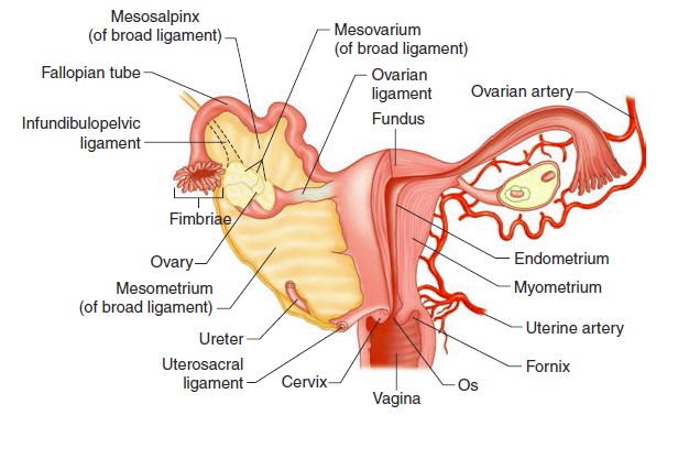 |
| Posterior view of female reproductive system. |
2) Accessory organs:
consists of:
1) The fallopian tubes (uterine tubes
or oviducts),
2) Uterus,
3) Vagina.
 |
| Female reproductive system. |
Location: In the female abdominal cavity. It extends from periphery of each ovary to uterus.
Structure: Pair of long, narrow ducts.
Function:
Function:
- Transportation of male sperm cells to the egg.
- Provides suitable environment for fertilization.
- Transports fertilized egg (embryo) into the uterine wall from ovary.
 |
| Fallopian tube of ovary. |
Size:
- 10-13 cm (4-5 inches) long.
- 0.5-1.2 cm (0.2-0.6 inch) in diameter.
Fallopian tube consists
of:
- Infundibulum,
- Ampulla,
- Isthmus.
Infundibulum:
Shape: funnel shape.
The edges of infundibulum have finger like projections called fimbriae (helps in collection of ova after ovulation).
Location: Proximal part of the fallopian tube.
Ampulla:
- Located next to infundibulum.
- It is wider central portion.
- It is the last part of the oviduct
Structure: It is short and thick-walled
structure.
Function: connects ampulla and infundibulum to uterus.
Size: 2 cm (0.8 inch) long.
2. Uterus (womb):
- It is hollow,
- Thick walled,
- Muscular,
- Highly vascular,
- Inverted peer shaped structure.
- It is the secondary sex organ,
- It provides mechanical support, nutritional support and waste removal for the developing embryo (weeks 1-8) and foetus (from week 9 until delivery).
- Responsible for the maintenance and transportation of the gametes,
- It has the capability to accommodate a growing foetus.
Location: Between urinary bladder and rectum, lying in pelvic cavity.
Uterus portion is covered by:
a) Fundus,
a) Fundus,
It is the
top rounded portion of the uterus, above the entry point of the uterine tubes.
b) Body:
b) Body:
It is the
usual site for implantation of the blastocyst.
c) Cervix:
The wall of uterus consists of:
c) Cervix:
- The uterus opens into the vagina through a narrow cervix.
- It is the lower part of the uterus. It is cylindrical or conical in shape.
- This part is structurally and functionally different to the rest of uterus.
- The cavity of the cervix is called the cervical canal.
- The cervical canal communicates with the vagina through external orifice and with uterus through internal orifice.
- The cervical canal along with the vagina forms the birth canal.
The wall of uterus consists of:
Perimetrium:
- It is the outermost thin membranous serous layer.
- It is the thick muscular middle layer.
- It exhibits strong contractions during parturition.
- It is the inner glandular layer.
- It undergoes cyclic changes during menstrual cycle.
- The female uterus contains one of the strongest muscles of human body.
- The female uterus is normally about 3 inches long and 2 inches wide but can expand 20 times during pregnancy.
- It is the large fibromuscular tube.
- It extends from cervix to the exterior.
- It accommodates the male penis during sexual intercourse.
Function: For copulation.
The female reproductive structure that lie external to the vagina are called as the external genitalia.
Structure: Inverted triangular area of fatty tissue. Extends from the pubic hairline. It is a fatty tissue, lies over the pubic symphysis.
It contains:
Location: Posterior to right and left openings vagina.
Location: On the anterior wall of vagina and around the lower end of the urethra.
Contains: Numerous microanatomical structures in common with prostate glands such as secretory cells.
3) External genitalia or vulva:
The female reproductive structure that lie external to the vagina are called as the external genitalia.
The
external genitalia comprising of:
1. Mons pubis:
- Mons pubis,
- Labia majora,
- Labia minora,
- Hymen,
- Clitoris,
- Bartholin’s glands, and
- Skene’s glands.
 |
| Vulva of female reproductive system. |
1. Mons pubis:
Structure: Inverted triangular area of fatty tissue. Extends from the pubic hairline. It is a fatty tissue, lies over the pubic symphysis.
Covered by: Hair follicles on the top of pubic bone.
2. Labia majora:
2. Labia majora:
- They are fleshy folds of tissue also known as “Major lips” or “Large lips”.
- They are homologous to scrotum in males (it is derived embryologically from the same tissue).
It composed of: Subcutaneous adipose tissue and fat.
Function: It encloses and protects other external reproductive organs.
It contains:
- Sweat and oil secreting glands,
- Fat,
- Pubic hair follicles, and
- Fewer touch and pressure receptors. In the surface the skin it contains squamous epithelium.
- Also known as “Minor lips” or “Small lips”.
Size: 3-4 cm long, 2 inches wide approximately.
Location: Underlying the labia majora, it extends anteriorly from clitoris to the
fourchette (a small fold of membrane connects labia minora in the posterior
part of vulva) posteriorly.
Labia minora contains: Rich in sebaceous glands, connective tissues
and vascular erectile tissue, with a considerable number of sensory nerve
endings and receptors.
Color:
Ranging from light pink to brownish black in colour in
different individuals.
4. Clitoris:
4. Clitoris:
- It is an erectile structure.
- It is homologous to male penis.
Clitoris formed by: Two corpora cavernosa and the glans, formed by
prepuce. It is rich in sensory receptors.
Visible portion: only a fifth (or less portion) is visible (glans)
while the rest is hidden under the skin. The glans is the externally visible portion
of the clitoris.
Size:
Average length is about 1-1.5 cm (0.5 inch) and 0.5 cm in
diameter. There is however, considerable variation in clitoral size.
Clitoris glans covered by: Partially by clitoral
prepuce, which is homologous to a similar structure covering the glans of the
penis.
5. Hymen:
5. Hymen:
- It is a thin ring of tissue.
- It is a septum of mucous membrane, which varies in shape greatly.
- It is usually crescentic or circular in virgins and is ruptured during sexual intercourse.
- It forms the part of vulva.
- Hymen is rich in nerve endings.
Variation: It ranges from
thin to thick, stretchy and may be rigid, may also be completely absent. An imperforate
hymen occurs in 1-2 out of 1000 infants.
Appearance: Normal appearance
of hymen is crescent shaped, although many shapes are possible. Due to secretion
of oestrogen hormone in puberty it become elastic and change in appearance.
Function: The external opening of the vagina
is partially closed by this hymen. The hymen is often torn during the first
coitus (physical union). It may also
remain intact in some women. It can be torn or stretched due to a sudden fall
or jolt and also during strenuous physical activities such as horseback riding,
cycling, etc., and therefore cannot be considered as an indicator of a woman’s
virginity.
6. The Bartholin’s glands (the Greater vestibular glands):
6. The Bartholin’s glands (the Greater vestibular glands):
Location: Posterior to right and left openings vagina.
Secretion: Mucous.
Function: To lubricate the vagina and are homologous to
bulbourethral glands of male.
7. Skene’s glands:
 |
| Image shows Bartholin's glands. |
7. Skene’s glands:
Location: On the anterior wall of vagina and around the lower end of the urethra.
Contains: Numerous microanatomical structures in common with prostate glands such as secretory cells.
Secretion: A lubricating fluid, homologous to the
prostate gland of males.
Mammary glands:
 |
| Image showing Skene's glands. |
Mammary glands:
- These glands are present in both males and females.
- They are modified sweat glands.
- It is rudimentary in males and functional in females.
It contains
variable quantities of fat with a median nipple and a glandular
tissue.
↓
The nipple
is surrounded by a pigmented area called the areola. Areolar glands or sebaceous
glands found on the skin to reduce the cracking of the skin of the nipple.
↓
Each
mammary gland consists of 2-25 lobes, and they are separated by connective
tissues and fats. Each lobe is made up of lobules.
↓
Lobules of
each lobe contains alveoli or acini lined by epithelial cells.
↓
The cells
of alveoli secrete milk.
↓
The alveoli
open into mammary tubules.
↓
The tubules
of each lobe join to form a mammary duct.
↓
Several
mammary ducts join to form a wider mammary ampulla.
↓
The ampulla
is connected to the lactiferous duct in the nipple.
↓
Under the
nipple, each lactiferous duct expands to form the lactiferous sinus.
↓
The
lactiferous sinus serves as a reservoir of milk.
↓
Each
lactiferous duct opens separately by a minute pore on the surface of the nipple.
The normal
development of breast begins at puberty and progress with changes during
each menstrual cycle. The glandular structure in non-pregnant women is
largely underdeveloped and the breast size is largely due to amount of fat
deposits. The size of breast does not have an influence on the efficiency of
lactation.
More to know:
 |
| Mammary glands-cross sectional view. |


Post a Comment
Post a Comment